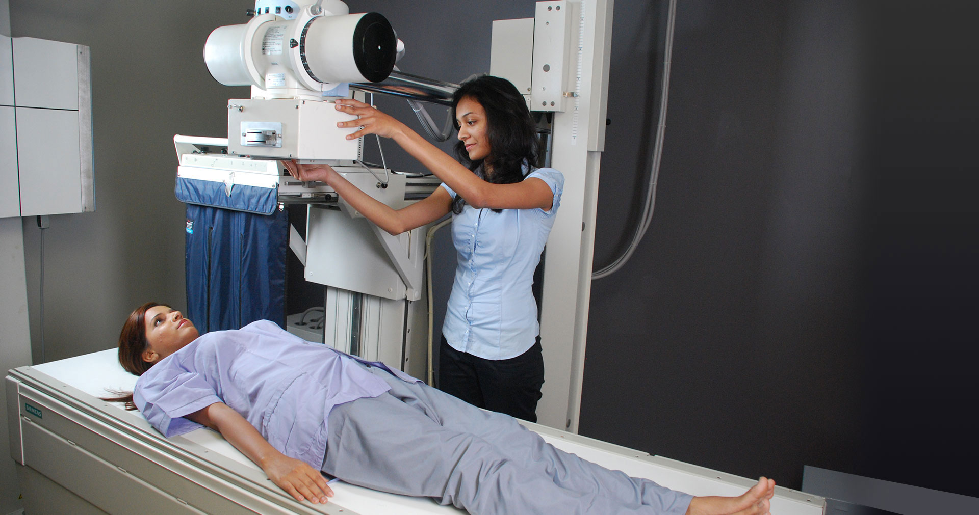
X-rays offer a fascinating glimpse inside the human body. This guide explores how these powerful energy beams help medical professionals diagnose everything from broken bones to tumors, delving into the different types of X-rays, their mechanisms, and what you can expect during a procedure. Discover how this revolutionary technology has transformed healthcare.
Decoding X-rays: How They Work
X-rays, a form of electromagnetic radiation similar to light but with significantly higher energy, are a cornerstone of modern medical imaging. When directed at the body, these rays penetrate different tissues at varying rates. Denser materials like bone absorb more X-rays, while softer tissues like muscle and skin allow more to pass through. This differential absorption creates a shadow-like image on a detector or film, revealing the underlying structures.
What Can X-rays Reveal?
X-rays offer valuable insights into various bodily systems, including:
-
Bones and Joints: X-rays excel at examining the skeletal system, revealing fractures, arthritis, bone abnormalities, and even herniated discs.
-
Digestive System: While other imaging techniques offer greater detail, X-rays can sometimes detect issues like kidney stones and certain blockages.
-
Respiratory System: X-rays are often the first step in diagnosing lung problems such as pneumonia and lung cancer.
-
Infections: X-rays can occasionally pinpoint the location of infections like abscesses.
-
Foreign Objects: From swallowed coins to embedded splinters, X-rays can locate foreign objects, guiding their removal.
Exploring Different X-ray Types
Just as different cameras serve specific purposes, various X-ray types exist:
| Type of X-ray | What it Examines |
|---|---|
| Abdominal X-ray | Abdominal organs (stomach, intestines, liver, kidneys) |
| Bone X-ray | Bones (fractures, arthritis, other conditions) |
| Chest X-ray | Heart, lungs, and surrounding chest structures |
| Dental X-ray | Teeth and jaw (cavities, infections) |
| Fluoroscopy | Real-time, continuous X-ray imaging during procedures |
Understanding the X-ray Procedure
An X-ray is typically quick and painless. Here’s what you might expect:
-
Preparation: You may need to remove clothing and jewelry in the target area. Contrast solutions or injections may be used to highlight specific tissues.
-
Positioning: A technician will carefully position you to ensure the correct body part is imaged.
-
Imaging: The X-ray machine briefly emits X-rays, creating the image. This is painless.
-
Waiting: Images are processed quickly, and a radiologist reviews them, sending a report to your physician. When thinking about a root canal, one of the most important questions is, how long does how long does a root canal last without a crown last without a crown? If you’re curious about the longevity and durability of a root canal, it’s a question that you’ll want to explore in more detail.
Assessing the Risks of X-rays
While valuable, X-rays have potential risks:
-
Radiation Exposure: X-rays involve a small dose of ionizing radiation, carrying a slight, theoretical risk of cell damage and a potentially increased cancer risk over a lifetime. However, the diagnostic benefits typically outweigh this minimal risk. Experts continually work to minimize exposure during procedures.
-
Pregnancy: Inform your doctor if you are or might be pregnant. While the risk from a single X-ray is generally low, avoiding unnecessary exposure during pregnancy is recommended.
-
Contrast Materials: Contrast materials (iodine, barium) can sometimes trigger allergic reactions. Inform your doctor about any allergies.
The Future of X-ray Imaging
Ongoing research aims to improve image quality while minimizing radiation exposure. Scientists are exploring techniques for more precise X-ray targeting, promising safer and more effective diagnostics.
Why Is It Called an X-Ray? Decoding the Mystery of the “X”
The “X” in X-ray stands for “unknown.” In 1895, physicist Wilhelm Conrad Röntgen discovered these rays while experimenting with cathode ray tubes. He observed a fluorescent glow, even when the tube was covered, suggesting an unknown type of radiation. Following scientific convention, he labeled this enigmatic radiation “X-radiation.” While the technical term remains “X-radiation,” the shortened, more pronounceable “X-ray” evolved through common usage and has become the standard term.
Röntgen’s Discovery: A Scientific Breakthrough
Röntgen’s serendipitous discovery revolutionized medicine. His first medical X-ray, an image of his wife’s hand, showcased the technique’s potential for non-invasive visualization of internal structures. This breakthrough paved the way for the development of medical imaging as we know it today.
X-rays: Properties and Applications
X-rays are a form of electromagnetic radiation with short wavelengths and high energy. This high energy enables X-rays to penetrate materials opaque to visible light. While X-rays are most commonly associated with medical imaging, they also have applications in security screening, materials analysis, and even art restoration. Current research continues to explore X-ray properties, refining techniques for improved image resolution and targeted applications, minimizing risks, and expanding possibilities within medicine and beyond.
- Glass Storage Containers With Glass Lids That Lock for Freshness - February 2, 2026
- Locking Glass Food Storage Containers for Organized and Fresh Meals - February 1, 2026
- Borosilicate Glass Food Storage for Freshness and Organization - January 31, 2026


