Dealing with nagging pain from bone spurs? This guide explains bone spurs (exostosis), what causes them, and how they’re treated. We’ll cover everything from simple pain management to surgery (exostectomy), helping you understand your options and make informed decisions. For more information on bone grafting procedures, see this helpful resource on bone graft costs. We’ll walk you through what to expect during and after surgery, including recovery and long-term care. Whether you’re considering surgery or trying to find relief without it, this guide will help you navigate the process and find the best path to pain relief.
Understanding Exostoses: Removing Those Pesky Bone Spurs
Bone spurs, also known as exostoses (singular: exostosis), are benign bony growths that develop on the surface of bones. They’re essentially extra bits of bone that grow where they shouldn’t. While often harmless, these bony protrusions can cause pain and discomfort, especially when located near joints or impinging on nerves. Think of them as the bone equivalent of a callous on your heel – annoying, and sometimes painful, but usually not a major health threat unless they interfere with movement or cause significant pain. They can show up anywhere, but common spots include your feet, ankles, knees, hips, spine, fingers, toes, and ear canals. It is estimated that exostoses affect approximately 6.5% of the global population, with prevalence varying based on age, activity level, and underlying medical conditions.
What Causes These Bony Outgrowths?: Examining Exostosis Etiology
While the exact cause of exostosis formation is not always known (often referred to as idiopathic), several factors can contribute to their development. These include:
- Injury: Trauma to a bone can stimulate bone growth and lead to the formation of a spur.
- Repetitive Stress: Activities that involve repeated stress on joints, such as running, jumping, or certain sports, can contribute to exostosis.
- Osteoarthritis: The wear and tear on joints associated with osteoarthritis can lead to the formation of bone spurs near the affected joint, as the body attempts to stabilize the area.
- Genetics: A family history of exostoses may increase your likelihood of developing them. Some conditions like Hereditary Multiple Exostoses (HME) are directly linked to genetic mutations affecting bone growth.
- Age: Bone spurs are more common in older adults due to the cumulative effects of joint wear and tear.
- Poor Posture: Long term poor posture can lead to uneven weight distribution and extra stress on certain joints, potentially initiating bone spur development.
- Underlying Conditions: Conditions such as spinal stenosis can also cause extra bone growth.
Children and adolescents, due to their rapid bone growth, are also slightly more susceptible, especially to conditions like osteochondromas, a type of exostosis that develops near growth plates.
Noticeable Symptoms (or Lack Thereof): Clinical Presentation
Many people with bone spurs experience no symptoms at all. These growths are often discovered incidentally during X-rays or other imaging tests performed for unrelated reasons. However, when symptoms do arise, they can vary depending on the location and size of the exostosis. Common symptoms include:
- Pain: Pain is the most common complaint, ranging from a dull ache to a sharp, stabbing sensation. The pain may worsen with activity or pressure on the affected area.
- Stiffness: Exostoses near joints can restrict movement and cause stiffness, making it difficult to perform certain activities.
- Limited Range of Motion: The bony growth can physically block the normal range of motion of a joint.
- Numbness or Tingling: If the exostosis presses on a nerve, it can cause numbness, tingling, or weakness in the affected area.
- Visible Bump: In some cases, you may be able to feel or see a bony bump under the skin.
- Inflammation: The area around the exostosis may become inflamed, causing swelling, redness, and warmth.
- Specific Symptoms Based on Location:
- Haglund’s Deformity: (heel spur) causes pain at the back of the heel, especially when wearing shoes.
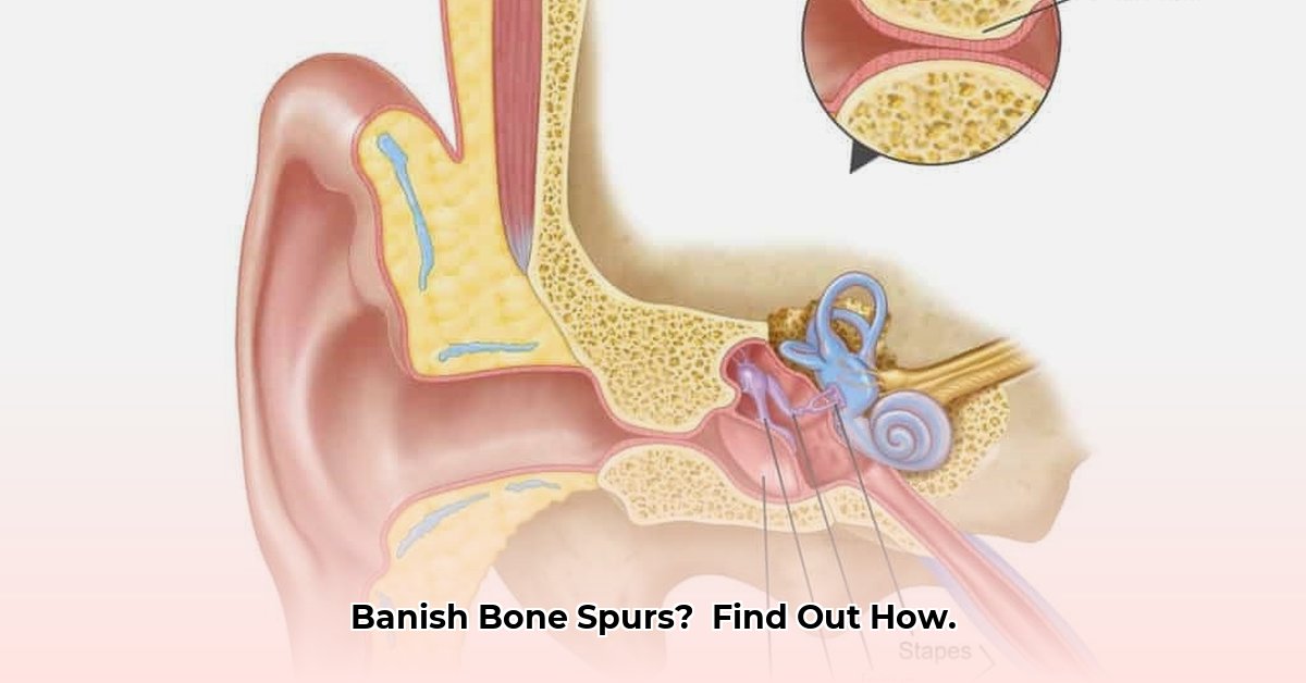
- Surfer’s Ear: (ear canal growth) can lead to hearing loss, ear infections, and earwax impaction.
- Hallux Rigidus: (big toe spur) limits movement of the big toe, causing pain and difficulty walking.
Diagnosis: Getting to the Root of the Problem with Imaging Techniques
Diagnosing an exostosis typically involves a combination of a physical examination and imaging tests.
- Physical Examination: Your doctor will begin by examining the affected area, feeling for any bony bumps or tenderness. They will also assess your range of motion and ask about your symptoms and medical history.
- Imaging Tests:
- X-rays: X-rays are usually the first imaging test ordered. They can clearly show the presence of bone spurs and help determine their size and location.
- CT Scans: Computed tomography (CT) scans provide more detailed images of the bone and surrounding tissues. They can be helpful in complex cases or when the exostosis is located in a difficult-to-visualize area.
- MRI: Magnetic resonance imaging (MRI) can help visualize soft tissues, such as nerves and tendons, and can be useful in determining if the exostosis is affecting these structures. Bone spurs are typically low signal on all MRI sequences
- Ultrasound: In some cases, ultrasound may be used to evaluate soft tissues around the bone spur.
The imaging tests are crucial for confirming the diagnosis, determining the exact size and location of the spur, and, importantly, ruling out more serious conditions like cancerous bone tumors or other bone abnormalities.
Treatment: More Than Just Pain Relief – Conservative and Surgical Treatment Options
Treatment for exostoses depends on the severity of symptoms and the impact on your daily life. Mild cases may not require any treatment at all, while more severe cases may benefit from conservative therapies or surgery.
Conservative Treatment Options:
- Pain Relievers: Over-the-counter pain relievers such as ibuprofen (Advil, Motrin) and naproxen (Aleve) can help reduce pain and inflammation. In some cases, your doctor may prescribe stronger pain medications.
- Rest: Avoiding activities that aggravate your symptoms can help reduce pain and inflammation.
- Ice: Applying ice to the affected area for 15-20 minutes at a time, several times a day, can help reduce swelling and pain.
- Physical Therapy: Physical therapy can help improve joint flexibility, strength, and range of motion. A physical therapist can also teach you exercises to help manage your symptoms.
- Orthotics: Shoe inserts or orthotics can help support and cushion the foot, reducing pressure on bone spurs in the foot or ankle.
- Lifestyle Modifications: Weight loss, improved posture, and ergonomic adjustments can help reduce stress on joints and prevent further exostosis growth.
Surgical Treatment Options:
If conservative treatments are ineffective or if the bone spur significantly limits joint function, surgery (exostectomy) may be recommended.
Exostectomy: Surgical Removal of Bone Spurs – A Detailed Look
Exostectomy is a surgical procedure to remove a bone spur. The procedure involves making an incision over the exostosis, carefully dissecting through surrounding tissues, and removing the extra bone using specialized instruments, such as osteotomes or burrs. The goal is to alleviate pain and restore normal joint function.
The Exostectomy Procedure: A Step-by-Step Look
- Anesthesia: You will receive either local anesthesia (numbing the area around the exostosis) or general anesthesia (putting you to sleep) depending on the location and complexity of the surgery, and your surgeon’s preference. Regional anesthesia is also common, especially for lower extremity exostectomy surgery.
- Incision: The surgeon makes a small incision directly over the bone spur. The location and size of the incision will vary depending on the location and size of the exostosis.
- Spur Removal: Using specialized surgical instruments, the surgeon carefully removes the extra bone, taking care to avoid damaging surrounding tissues, such as nerves, blood vessels, and tendons.
- Closure: Once the bone spur has been removed, the incision is closed with sutures or staples. A sterile dressing is applied to protect the wound.
After the Surgery: Recovery and Rehabilitation – What to Expect
Recovery after exostectomy surgery varies depending on the location and extent of the procedure. However, some general guidelines include:
- Pain Management: Pain medication will be prescribed to manage post-operative pain.
- Immobilization: You may need to wear a splint, cast, or brace to protect the area and promote healing. The duration of immobilization will depend on the location of the surgery.
- Weight-Bearing Restrictions: You may need to limit weight-bearing on the affected limb for a period of time. Your surgeon will provide specific instructions based on your individual needs.
- Wound Care: Keep the incision clean and dry. Follow your surgeon’s instructions for dressing changes.
- Physical Therapy: Physical therapy is often recommended to regain strength, flexibility, and range of motion in the affected joint.
- Elevation: Elevating the surgical site, especially in the first few days after surgery, can help reduce

- Easy Meal Prep Ideas for College Students for a Budget - March 16, 2026
- Easy College Meal Prep Ideas to Save Time and Money - March 15, 2026
- Easy College Student Meal Prep Ideas for Healthy Budget-Friendly Meals - March 14, 2026